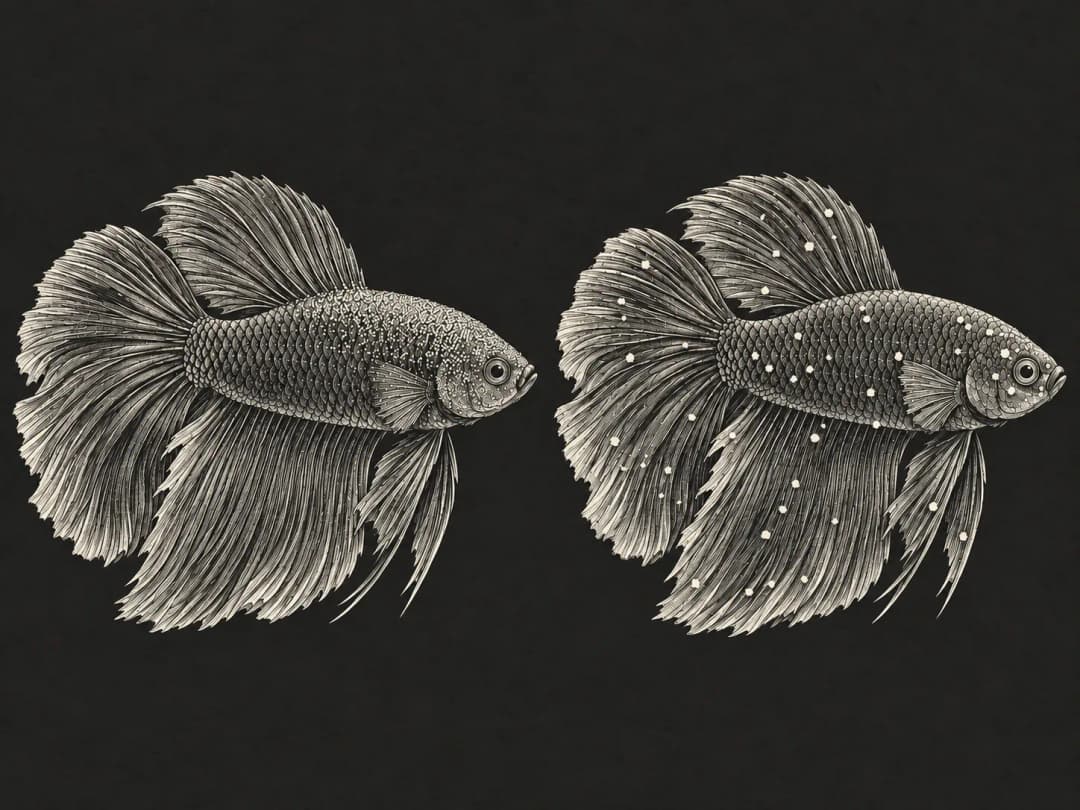
Fine golden or rust-colored dust on the body of a freshwater fish may be linked to velvet disease; but some infections are concentrated mainly in the gills, where the typical gold dust can't be seen with the naked eye. Changes in breathing, behavior and appetite deserve just as much attention.[1][2]
In freshwater, the pathogens are mainly parasitic dinoflagellates of the genus Piscinoodinium, with P. pillulare among the common species.[1][6] Marine velvet is typified by Amyloodinium ocellatum, and treatment information for the two can't be used interchangeably.[1]
What to check first if you suspect velvet
Measure water temperature, pH, ammonia and nitrite, and check aeration and filtration. Watch for flashing, clamped fins, refusal to eat and rapid breathing, and note whether new fish were added recently or water quality changed.
If young fish are affected, several fish become abnormal within a short time, or fish are breathing rapidly with no obvious lesions, contact a veterinarian as soon as possible. Choosing a medication based only on the color of the dust makes it easy to miss gill infections or misjudge the cause.
The gold-dust look and gill infections you can't see
The parasitic stage carries pigment and attaches to the skin, fins or gills, and heavy infections can give the fish a velvety sheen. When the gills are affected, pathology studies in ornamental armored catfishes (Corydoras, Brochis) have found epithelial hyperplasia and fusion of the gill lamellae, which may reduce the effective area for gas exchange.[1][3]
Affected fish may rub against objects, swim weakly, refuse food or stay near the surface. These signs can also be seen with low oxygen and other diseases, so water quality results need to be interpreted together with microscopy. Young fish are often more vulnerable to this pathogen.[2]
How to tell velvet from ich
| Feature | Freshwater ich | Freshwater velvet |
|---|---|---|
| Pathogen | Ciliate Ichthyophthirius multifiliis | Dinoflagellate Piscinoodinium |
| Typical appearance | Distinct, salt-grain-like white spots | Finer golden, rust-colored or grayish-white dust, which may look like a film |
| Main sites | Skin, fins, gills | Skin, fins, gills |
| No spots on the body | Gill infection still possible | Gill infection also possible |
| How to confirm | Wet-mount microscopy of the affected tissue | Wet-mount microscopy of the affected tissue |
Table compiled from fish parasite resources.[1][2][4] Under the microscope, the parasitic stage of Piscinoodinium is usually oval, amber, granular inside and doesn't move freely;[2] ich parasites have different movement and cell features.[8] These exams should be done by professionals.
Why the life cycle leads to repeat outbreaks
The life cycle includes a parasitic stage on the fish, an encysted stage after it leaves the fish, and free-swimming dinospores in the water. Mature parasites leave the fish and divide inside the cyst to produce new infective stages; when the dust on the fish fades, the pathogen may still be present in the environment.[1][5]
An early UF/IFAS guide to freshwater parasites notes that at about 23–25°C the life cycle can take 10 to 14 days, and possibly longer at lower temperatures. This is a reference range from a specific source, not a fixed timeline for every aquarium, and it shouldn't be used to work out dosing frequency on your own.[5]
Cysts are more resistant to chemical treatment.[5] Treatment plans should be based on the confirmed pathogen, the fish species and the product used; drug sensitivity and treatment-course data for marine Amyloodinium can't be used directly as a prescription for freshwater Piscinoodinium.
Do chloroplasts mean keeping the tank dark will cure it?
The parasitic stage of Piscinoodinium has pigment and can photosynthesize, and some case studies have also observed chloroplasts.[1][6] But this biological feature doesn't directly prove that darkening a home aquarium alone will clear the infection, and darkening certainly can't replace confirming the pathogen and completing treatment.
Likewise, adjusting the water temperature isn't a universal approach that suits every species, and you shouldn't change it on your own. Any environmental adjustment needs to take the fish's tolerance and breathing into account and be discussed with a veterinarian.
Reduce the risk of bringing in pathogens and spreading infection
Quarantine new fish in a separate system first, don't pour shipping water into the tank, and keep nets and buckets separate. Maintain appropriate stocking density, good aeration and water quality, remove excess organic debris and reduce stress on the fish.[7]
Velvet disease can cause heavy losses.[1][6] Infection or mortality rates from published case reports can't be taken directly as the risk in a home aquarium; how urgent it is depends on the fish's actual breathing, the course of illness and how many fish are affected.
Frequently asked questions
If shining a flashlight reveals gold dust, is that enough to diagnose it?
No. Light can help you look at the body surface, but natural reflections, mucus and other attached material can be confusing, so a diagnosis still requires professional examination.
I don't see any gold dust, but my young fish keep dying. What should I do?
First rule out water quality and dissolved oxygen problems, then have a veterinarian check the gills and other causes; the absence of typical gold dust doesn't rule out velvet disease.[1][2]
If there's less dust, has the fish recovered?
The dust may fade as parasites leave the fish, while the environmental stages can keep developing. Whether treatment is complete depends on the fish's overall condition and the treatment course.
If medication is needed, use a legally registered veterinary drug that's appropriate for the case, following the label and your veterinarian's instructions.
Further reading
- What to Do When Your Fish Is Sick – Reading Aquarium Fish Symptoms and What to Do First
- Ammonia and Nitrite Poisoning – Fish Gasping at the Surface and How to Read Water Test Kits
- How to Quarantine New Fish – Isolating for at Least 30 Days and Acclimating Fish to the Tank
- White Spot Disease (Ich) in Freshwater Fish – The Ichthyophthirius Life Cycle and Why It Keeps Coming Back
References
- Petty BD, Francis-Floyd R, Yanong RPE. Parasitic Diseases of Fish. Merck Veterinary Manual, revised 2022.
- Pouder DB, Curtis EW, Yanong RPE. Common Freshwater Fish Parasites Pictorial Guide: Dinoflagellates, Coccidia, Microsporidians and Myxozoans. UF/IFAS, FA110, 2005.
- Ferraz E, Sommerville C. Pathology of Piscinoodinium sp. (Protozoa: Dinoflagellida), parasites of the ornamental freshwater catfishes Corydoras spp. and Brochis splendens (Pisces: Callichthyidae). Diseases of Aquatic Organisms 33, 43–49, 1998. doi:10.3354/dao033043.
- Francis-Floyd R, Yanong RPE, Pouder DB. Ichthyophthirius multifiliis (White Spot) Infections in Fish. UF/IFAS, CIR920/FA006, 1991 (revised 2026).
- Klinger RE, Francis-Floyd R. Introduction to Freshwater Fish Parasites. UF/IFAS, CIR716, 1987 (reviewed 2013).
- Shaharom-Harrison FM et al. Epizootics of Malaysian cultured freshwater pond fishes by Piscinoodinium pillulare (Schaperclaus 1954) Lom 1981. Aquaculture 86, 127–138, 1990. doi:10.1016/0044-8486(90)90107-X.
- Yanong RPE, Erlacher-Reid C. Biosecurity in Aquaculture Part 1 An Overview. SRAC Publication 4707, 2012.
- Pouder DB, Curtis EW, Yanong RPE. Common Freshwater Fish Parasites Pictorial Guide: Motile Ciliates. UF/IFAS, FA108, 2005.