
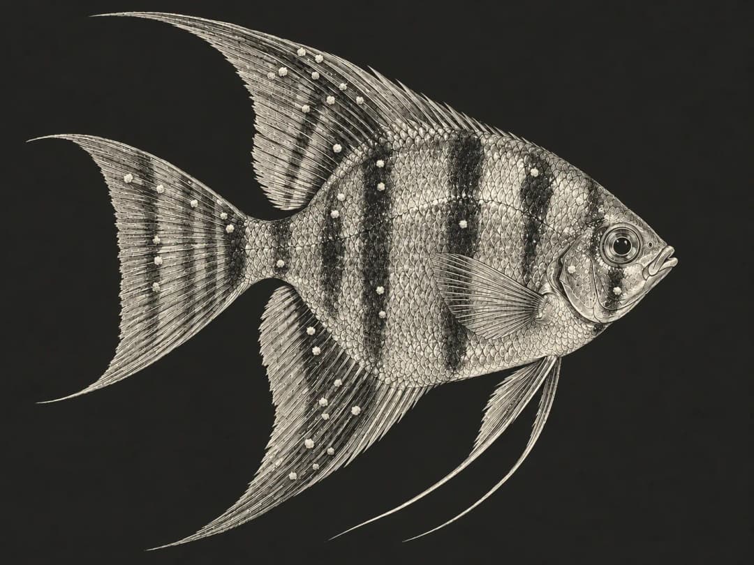
When salt-grain-like white spots appear on a freshwater fish, ich is an important differential, but it still needs to be confirmed under a microscope. The spots fading may just mean mature parasites have left the fish; cysts on the tank bottom may still be releasing larvae, so you can't stop treatment based on appearance alone.[1]
This article covers the freshwater ich parasite Ichthyophthirius multifiliis. White spot disease in marine fish is caused by a different ciliate, Cryptocaryon irritans, and the life cycle and treatment timeline of one can't be applied to the other.[2] White spot syndrome virus in shrimp is a virus that infects shrimp and other decapod crustaceans, and it's also different from ich in fish.[6]
What to do first when you see white spots
Measure water temperature, pH, ammonia and nitrite, and check aeration and filtration. Note how many fish are affected, their breathing and appetite, and whether new fish were added recently or the temperature changed sharply. If several fish are breathing rapidly, fish keep dying without any white spots, or the spots increase rapidly, contact a veterinarian as soon as possible.
Don't decide treatment is finished just because "the spots are gone," and don't raise the temperature on your own. Once the pathogen is confirmed, plan a full treatment course based on the species, water temperature and the product label; frequently switching medications or treating only once may fail to cover parasites at different stages.[1][2]
Symptoms beyond white spots
Affected fish may flash, clamp their fins, refuse food, become lethargic, darken in color or produce excess mucus.[1][2] Ich can parasitize the skin, fins and gills; if the infection is concentrated in the gills, there may be no obvious white spots on the body even though breathing is already affected.[1]
Microscopy examines the affected tissue and identifies the parasite by its movement and cell features. The size of the spots, whether they're raised or where they're located can't reliably rule out other parasites and lesions on their own.[1][2]
Why the ich life cycle affects the timing of treatment
| Stage | Location | Relevance to treatment |
|---|---|---|
| Parasitic stage (trophont) | Inside the skin or gill epithelium of the fish, feeding and growing | Protected by the fish's tissue and mucus, it's usually hard to eliminate with chemical treatments in the water. |
| Cyst stage (tomont) | Attached to surfaces in the environment after leaving the fish, dividing inside the cyst | The cyst protects the parasites inside, which go on to release new batches of larvae. |
| Infective stage (theront) | Free-swimming in the water, searching for a fish host | This is the vulnerable stage that routine chemical treatments mainly target. |
Life cycle summarized from UF/IFAS.[1] A single cyst can produce large numbers of larvae, and once an infection is established, different stages are usually present at the same time. A single treatment can only affect the parasites exposed at that moment, and new larvae keep being released afterward, so repeated or continuous treatment needs to be planned according to conditions.
Fewer white spots and parasites leaving the fish may happen at the same time; this can simply be part of the life cycle and doesn't on its own prove the pathogen has been cleared.
Water temperature changes the length of the life cycle
UF/IFAS notes that at 24–26°C, one life cycle takes about three to six days; in cold water it may stretch to several weeks. These times vary with the isolate and environment and can only help you understand how fast the cycle runs; they can't be converted directly into a dosing interval for every medication.[1]
At lower water temperatures, the next batch of larvae may appear later and the total treatment period may be longer; at higher temperatures the cycle speeds up, but the fish's tolerance range and oxygen needs must also be considered.[1][2]
Can raising the temperature alone cure ich?
Raising the temperature shouldn't be treated as a reliable way to deal with ich. Two studies using isolates from rainbow trout showed that ich cysts could still develop and release larvae at 30°C, so it's unwise to assume that raising the temperature will break the life cycle.[3][4] Fish species also differ in their tolerance of high temperatures.
Raising the temperature changes the fish's metabolism and how much oxygen the water can hold. Whether to adjust the water temperature should be assessed by a veterinarian together with the species, the fish's breathing and the overall treatment plan, rather than following a fixed temperature from the internet. If medication is needed, use a legally registered veterinary drug that's appropriate for the case, following the label and your veterinarian's instructions.
How gravel cleaning and leaving the tank fishless can help
Removing debris and organic matter from the bottom can reduce the cysts attached in the environment and the pathogen load. Cleaning is a supporting measure and can't guarantee that siphoning alone will clear the infection; avoid over-disturbing sick fish or disrupting biological filtration while cleaning.[1][2]
Ich needs a fish host to complete its life cycle, so leaving the original system without fish can lower the risk of reinfection. But the fishless period must cover the environmental stages at the current water temperature; "larvae die quickly if they can't find a fish" can't be simplified into "an empty tank is safe after a day or two." Cold water in particular calls for a longer period.[1][2]
Lesions easily mistaken for white spots
| Possible look-alike | Limits of identification |
|---|---|
| Velvet disease | May look like fine dust or have a golden-rust color, but color alone can't confirm a diagnosis. |
| Lymphocystis disease | May appear as wart-like nodules; small early nodules can also be confusing. |
| Epistylis and other attached ciliates | Attached to the fish and growing in colonies, common in water with high organic levels, and may come with bacterial lesions; can't be told apart by appearance alone. |
Differences summarized from resources on parasites, sessile ciliates and lymphocystis disease.[2][5][7] If you're unsure, microscopy is more valuable than repeatedly switching treatments.
Prevention centers on new fish and cross-contamination
Quarantine new fish in a separate system first and use dedicated nets and buckets. Fish that have recovered from an infection may have partial immunity, or may carry a low-level infection without symptoms; "the older fish are fine" doesn't prove the system is free of ich.[1][2]
Reduce crowding, maintain water quality and avoid sharp temperature swings; when bringing in plants, substrate and other wet items, consider the pathogen risk as well. For the full isolation process, see "How to Quarantine New Fish".
Frequently asked questions
Can I stop medicating once the white spots are gone?
Don't use the disappearance of white spots as the only criterion. Whether the treatment course is complete should follow the label of the product used and your veterinarian's instructions, and the course should cover the pathogen at its different stages.[1]
Is it enough to isolate only the fish with white spots?
Not necessarily. The original system may already contain cysts and larvae, and other fish may have infections that aren't visible yet, so the whole connected system needs to be assessed.[1]
If a fish is gasping but has no white spots, can ich be ruled out?
No. The infection may be concentrated in the gills; also rule out low dissolved oxygen, water quality poisoning and other gill diseases.
Further reading
- What to Do When Your Fish Is Sick – Reading Aquarium Fish Symptoms and What to Do First
- Ammonia and Nitrite Poisoning – Fish Gasping at the Surface and How to Read Water Test Kits
- How to Quarantine New Fish – Isolating for at Least 30 Days and Acclimating Fish to the Tank
- Velvet Disease in Freshwater Fish – Gold-Dust Symptoms and How It Differs from Ich
References
- Francis-Floyd R, Yanong RPE, Pouder DB. Ichthyophthirius multifiliis (White Spot) Infections in Fish. UF/IFAS, CIR920/FA006, 1991 (revised 2026).
- Petty BD, Francis-Floyd R, Yanong RPE. Parasitic Diseases of Fish. Merck Veterinary Manual, revised 2022.
- Forwood JM, Harris JO, Landos M, Deveney MR. Life cycle and settlement of an Australian isolate of Ichthyophthirius multifiliis Fouquet, 1876 from rainbow trout. Folia Parasitologica 62, 013, 2015. doi:10.14411/fp.2015.013.
- Aihua L, Buchmann K. Temperature- and salinity-dependent development of a Nordic strain of Ichthyophthirius multifiliis from rainbow trout. Journal of Applied Ichthyology 17, 273–276, 2001. doi:10.1046/j.1439-0426.2001.00279.x.
- Yanong RPE. Lymphocystis Disease in Fish. UF/IFAS, FA181, 2010 (revised 2024).
- World Organisation for Animal Health (WOAH). Infection with white spot syndrome virus. Manual of Diagnostic Tests for Aquatic Animals, Chapter 2.2.8, 2023.
- Pouder DB, Curtis EW, Yanong RPE. Common Freshwater Fish Parasites Pictorial Guide: Sessile Ciliates. UF/IFAS, FA107, 2005.